Renal
Indications
· Colic
· Urinary Retention
· Pyelonephritis
· Question of Mass
· Trauma
Anatomy
R kidney is usually larger and lower than the left
Inferior poles are anterior and lateral to the superior poles
Technique
3.5 MHz unless thin
Right Kidney
Scan in sag at mid-clavicular line or coronal at mid ax (Point Marker towards post-ax). Start at costal margin and move down.
Left Kidney
Mid ax line. May need to go up on ribs. Scan may be aided by placing patient in right lateral recumbent
Findings
· R sided hydro during pregnancy is common and not pathologic
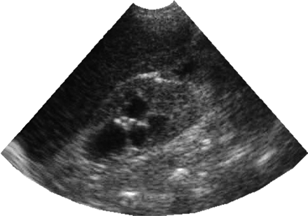
· Pyelo appears as normal kidney until abscess which appears as a round, hypoechoic mass
· Absence of ureteral jets by Doppler is 100% sensitive for obstruction
· Hydro can be masked by dehydration, and simulated by a full bladder. Give IV fluids before scanning
· Normal renal parenchyma is less echogenic than the liver
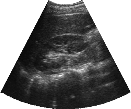
· Obstructive Uropathy
Hydronephrosis appears as echo-free areas in the sinus. Bladder volume can be measured with built-in calculators, but estimation is usually sufficient.
Variants
· Sonolucent pyramids-looks like hydro, but in the medulla, not the sinuses
· Duplication of collecting system-split in the sinus
· Ectopic Kidney
· Prostate Enlargement
| | |