http://www.upei.ca/~vca341/usphysics/img0.html
http://courses.washington.edu/radxphys/Lectures05-06/Ultrasound%20-%20Chapter%2016%20-%20Lecture%202%20-%20060302_files/frame.htm
http://courses.washington.edu/radxphys/PhysicsCourse.html
Physics
T=1/f
Hertz=cycles per second
1 million hertz=1MHz
Medical UTS=1-30 MHz
Audible sound = 20-20000 Hz
Impedance=C/density
Wave Length
(insert equation)
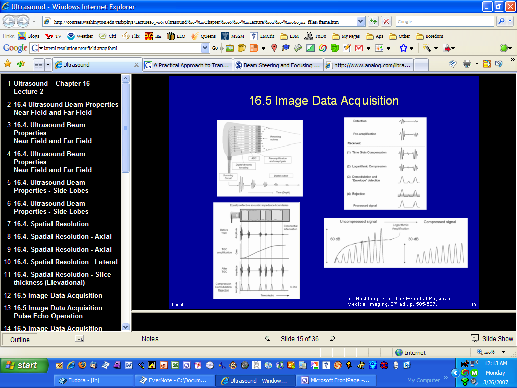
wave length is the distance between two peaks or valleys in the wave
it is determined by speed divided by frequency
For soft tissue,
use speed of sound as 1540 m/s
change it to mm
1.54 mm/f (MHz) gives the answer in mm
air and lung has the lowest impedance, bone has the highest
Reflection
Perpendicular Incidence
if materials with two different impedances are next to each other, some of the beams energy will be reflected and some will be transmitted.
Non-Perpendicular Sound Beam Incidence Can cause refraction, i.e. bending of the sound beam
Rayleigh Scatterers
structures smaller than the wavelength
they increase as frequency increases
Attenuation in tissue
caused by reflection and scattering and direct absorbtion
the higher the frequency, the higher the attenuation. almost proportional
beam characterisitcs
near field=rough waves
far field=smooth waves
with increasing frequencies, the near field length increases
beam divergence is less at higher frequencies
the diameter of the probe also increases the NFL
Ring-Down Artifact
wherever small bubbles or partial liquids exist
look like comet tails
Mirror Images
echo bounces off the bottom of the image hits an object and bounces back to the bottom
Doppler Spectral Mirroring
when gain is set too high or when angle is perpendicular to flow
Masses can show enhancement or shadowing
low attenuating masses generate large enhancement i.e. a cyst or fluid filled structure causes enhancement behind it
Refraction
at the interface of tissues with different speeds of sound
bone has major refraction problems
fat which is slower than tissue
Edge shadowing of structures like GB is another example
if a structure with a slower speed of sound is imaged everything distal to it will look further away
wavelength = c/f (just divide 1.54/freq)
Impedance=C/density
Intensity=mW/cm2
Incident Beam= 10 log (I2/I1)

Spatial resolution
axial and lateral resolution
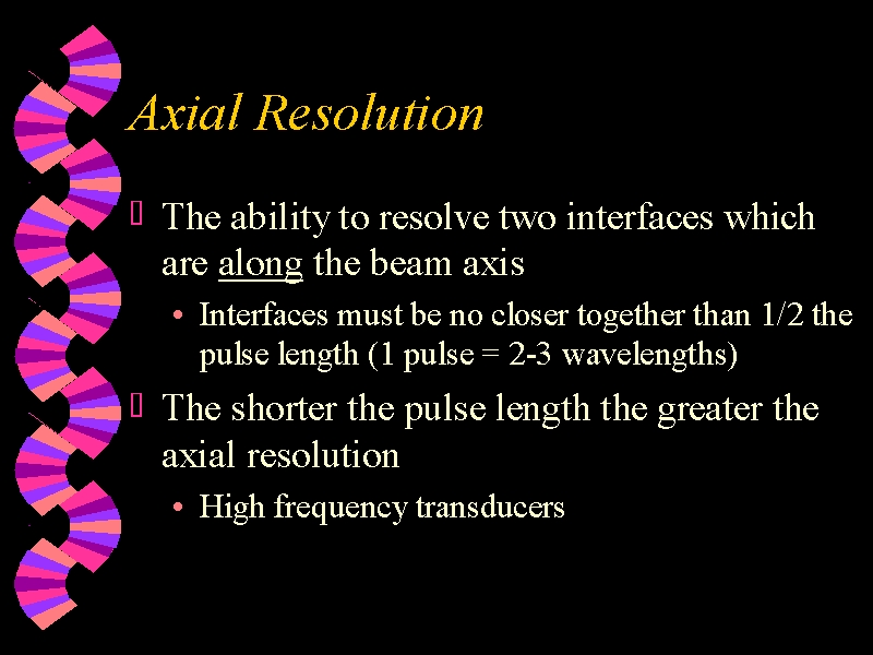
Axial Resolution
Longitudinal discrimination the minimum separation of two targets in tissue in a direction parallel to the beam which results in their being imaged as two distinct structures
Relates to pulse length (about 3 wavelengths)
Made better with higher frequency (b/c wavelengths are shorter)
Does not vary with depth
Axial resolution improves with increased damping, increased frequency, increased bandwidth, decreased pulse length
Transducers have a tendency to “ring” after being excited by an electrical impulse, creating an acoustic pulse which has an extended length in
Moreover, shortening the length of an ultrasound pulse while keeping the total energy of the pulse constant, results in a higher peak acoustic intensity. Thus a compromise is reached between the peak pressure to which tissue is exposed and the effective axial resolution of the ultrasound image.
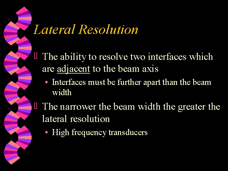
Lateral Resolution
relates to beam width
number of scan lines
wider transducer
higher frequency has a longer near zone and therefore narrower beam as long as area in focal zone
depth dependent
Shadowing of stones is lateral resolution
Near zone length is increased by increased frequency and wider transducer
Lateral resolution better with increased frequency and focusing (also by curving transducer)
focal zone=where beam is narrowest
fresNel=near
fraunhFer=far
focusing decreases bandwidth, improves lateral resolution not axial
focusing increases pulse length which hurts axial resolution
increased transducer diameter=increased near zone length=better LATERAL resolution
power does not affect it
receiver gain does not affect it
convex=wider image in near field and increased resolution at depth
Elevational Resolution
works just like lateral, relates to depth. has a focal zone as well
elevation=across the width of transducer=z thickness=slice thickness=mechanical focus by manufacturer
small cystic structures=elevational resolution
can cause pseudo-sludge b/c back of gb may wider beam than front
Temporal Resolution
frame rate
no phantom for this one
to improve temporal resolution decrease scan line density
frame rate=images per second 10-50 is the norm
scan depth is operator control which affects frame rate
decreasing focal zones increases temporal resolution as does increased prf
Contrast resolution
resolution of objects with similar reflective properties
contrast resolution-change of gray scale map

Frequency
all frequencies have identical transit times and sound propagation speeds
Propagation speed=speed of sound through substance, not user adjustable
Increased frequency=better spatial resolution and poorer penetration
increase frequency to see shadows because the stone must be wider than sound beam to see shadows
As you increase the frequency and focus the beam, the beam width narrows; therefore better axial resolution
shortest wavelength=highest frequency
As frequency increases, scattering, absorption, and attenuation all increase
2.5 Mhz to 5 Mhz probe, wavelength halves
Scattering intensity=frequency 4th power
If frequency is doubled, absorption is doubled
Resonance frequency=voltage frequency and thickness of an element
thin elements=high frequency
voltage of pulser determines final frequency
rate determines PRF
Power
increased power=increased penetration, acoustic power, brightness, and voltage
Power=Energy/Time
Decreasing Db
half value layer=decrease 3DB, shallower with higher frequency, point at which beam intensity is reduced by half
10 Db decrease is 10% of original
3 Db decrease is ½ of original
Propagation Speed
Tissue=1540 m/s or 1.54
Bone=4080
increased density causes decreased propagation speed
molecules oscillate (compressions and rarefactions) to propagate sound
rarefactions=low pressure/density formed during sound propagation
compression=elevated pressure during sound propagation
the stiffer the material, the quicker the sound
ONLY MEDIA determines the SPEED of SOUND
fat causes axial misrepresentation things look FURTHER AWAY, because fat is slower
Impedance
lung has highest rate of attenuation
fat has slowest propagation speed of tissue
impedance increases if density increases or speed increases and affected by stiffness, unaffected by frequency
air reflects all sound, 99.9% reflection coefficient
Gel reduces impedance difference between transducer and skin
unit of impedance=Rayl
Impedance=density(propagation speed)
Z=pc
Pulsed ultrasound-# of pulses to element per second=Pulse Repetition Frequency (PRF)
thickness of piezoelectric determines frequency
If number of cycles increase but wavelength stays the same, pulse duration is increased???
PRF=# of impulses to transducer/second
Period=time for one cycle
frequency=cycles per second
Period=1/frequency
milli=10 -3
micro=10 -6
absorption=sound converted to heat
transmission + reflection coefficient=100%
Reflection & Refraction
RBC=rayleigh scatterer
refraction=edge shadowing=different propagation speeds
reflection requires a difference in acoustic impedance
Specular reflections=renal capsule or diaphragm. LARGE SMOOTH INTERFACE
Specular reflection=crap image from oblique angles
Scattering=non-specular reflection-THIS IS WHAT ALLOWS IMAGING IN THE FIRST PLACE
interference=summation of waves
diffuse reflection=rough surface
diffraction=passage through aperture
Refraction described by Snell’s law=angle of sound c oblique interface and different speeds. Refraction
Attenuation
attenuation in soft tissue=0.5 dB/cm/MHz so as frequency goes up, attenuation goes up
have to double the depth b/c it is roundtrip
High attenuation (gallstone)=shadow
Low attenuation (bladder)=enhancement
½ power distance (1/2 value thickness) in cm water 380, blood 15, tissue 5, muscle 1, lung 0.05
Increased pressure = increased intensity
Absorption=sound to heat
Normal incidence=perpendicular incidence
Huygen’s instructive to deconstructive interference from each sound source
air is best reflector
Crystals & Elements
Curie=temperature point at which ceramics go piezo
crystal material=lead zirconate
aperture focusing=# of elements changed
Mechanical Sector
Mechanically steered
Mechanically focused
Linear
only one mechanical focus on width of beam
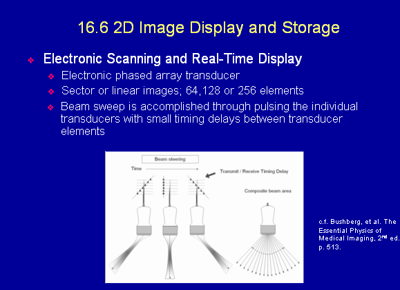
Phased array
Sector image, pointed top
Electronically Steered and focused
Annular array
mechanically steered, electrically focused
Beam is symmetrical about beam axis
Annular arrays are transducer assemblies with circular or ringlike elements, used to focus the beam. Annular arrays must be steered mechanically since they can only be fired in an outward-inward progression due to the rings. Annular arrays reduce section thickness artifacts
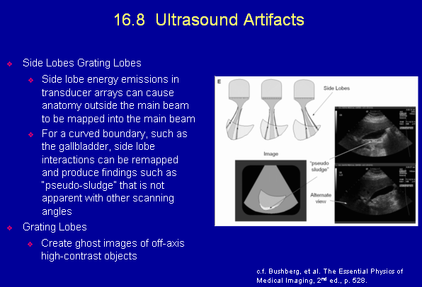
Side Lobes/Grating Lobes
Dynamic apodization=reduces side lobes makes all energy come from center of elements
subdicing=reduces grating lobes, breaking elements into sub-elements
Matching layer
reduces acoustic mismatch
matching layer should be 1/4 of wavelength
Focusing
dynamic receive focusing holds sound waves with others at same depth return
backing material
dampens ringing
dynamic damping-stop crystal from ringing after it rings
Gain
gain-volume knob of stereo
TGC-works on received echoes at depth
rejection-lowers electric noise, rejects low level echoes
the whole pulsed thing is to allow depth calculations
elements only fire about 1% of the time
reduce gain if background noise (i.e. not black background)
Processing
if it can be performed on a frozen image, it is POST PROCESSING
frame averaging-compares betweens frames and reduces random noise
tissue harmonics-improved contrast resolution, 2x transmitted frequency
Beam former apodization, beam steering, focusing aperture control?
interpolation-fills in skipped lines with decreased scan line density
pulser to beam former to receiver to memory to display
typical frame rate=10-50 Hz=10-50 frames per second
improved signal to noise=frame averaging
if only PRF is increased, frame rate will increase because it will take less time to fire all the pulses to make one frame. If too high, you get range ambiguity. New pulse fired before the first one returns
Signal to noise-system sensitivity; greater this ratio, the smaller the signal that can be differentiated
Rectification converts negative portion of signal to positive
Increasing dynamic range decreases image contrast because more levels to assign colors
Gain is at the receiver
range equation d=1/2 ct
3 DB decreased by 1/2
radio frequency to video=demodulater
AKA amplitude or envelope detection
transducer to electricity to acoustic pulses
duty factor is only time
increased PRF = increased duty factor
threshold is another name for rejection
read zoom-uses stored data
write zoom gets new data
scan converter-makes 2d image
Artifacts
Reverb-closely spaced reflections, like metal fragment
Pulse echo imaging
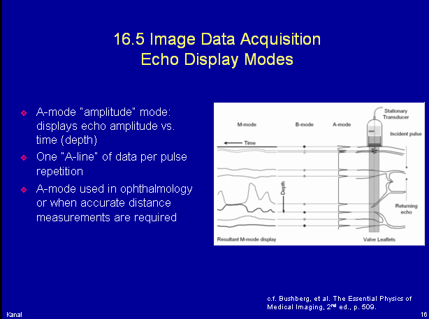
M-Mode
motion
depth of reflections with respect to time
m-mode=time, motion pattern
A-mode
amplitude
width of spike=strength of the echo
amplitude/distance (time) used in opthal
B-Mode
Brightness
Pixels
brightness level limited by bit depth or bits per pixel
numbers of shades of gray=contrast resolution, bits per pixel
television is 30 frames per second, 525 lines
digital scan converter = image memory storage area # of pixels in matrix=better spatial resolution
8 bit=1 byte=256 shades of gray
color needs 24 bis/pixel
if image is washed out, check processing equipment
memory to proportional voltages to brightness on monitor to digital to analog
Doppler
beat frequency=reference wave + reflected wave
continuous wave doppler-no max velocity
pulsed wave-max velocity
with pulse wave doppler, frequency shift > 1/2 PRF you get aliasing
to improve sensitivity to slow ???; decrease PRF as slow frequency=low freq shifts can also decrease wall filter and increase the doppler frequency
ensemble length=pulses per scan line
spectral broadening-fill in of spectral window associated with turbulent flow
indeterminate doppler angle-velocity estimate inaccurate
max frequency shift at 0° to flow; if angle is 90, no shift detected
determine direction, phase quadrature detection
Continuous wave Doppler
no range
flow towards=red=POSITIVE SHIFT
increased wall filter=reduced display of low frequency
Increased packet size=decreased frame rate, improved signal to noise
Fourier Analysis-used to perform spectral analysis for pulsed doppler
angle near 90°-you get spectral mirroring
aliasing top clipped off seen at bottom–fix by increased PRF, i.e. velocity scale, range, flow rate
doppler shift=difference between transmitted and received frequency
change F=2 V cos
smaller the angle, the larger the shift
Nyquist limit=aliasing frequency
reduce aliasing by increasing angle, lower zero baseline, increased PRF, decrease doppler frequency
aliasing=shift >1/2 PRF
color doppler uses autocorrelation
power doppler
encodes amplitude
does not use phase
angle does not matter
only strength of frequency
spectral broadening= turbulent flow
color gate=axial length of the sampling volume
spectral analysis-determines distribution and magnitude of frequency
to better image deep vessels, decrease doppler frequency
Image Features and Artifacts
partial volume artifact-from slice thickness that is too wide
ringdown=gas bubble
Quality Assurance
string phantom-doppler velocity
doppler flow phantom-velocity estimation, accuracy of flow directions
hydrophone-acoustic output level
sensitivity-ability to detect weak echoes
Closely spaced targets at various distances from the probe=AXIAL resolution
clear anechoic tubes oriented perpendicular =elevational resolution
width of point target=lateral resolution
adjust to maximum output and gain when testing
dead zone=distance from transducer to 1st echo
focal point=best lateral resolution
SMPTE-evaluates the display
Safety
1° C max increase in temperature
SATA=lowest for pulsed-wave field
increased focusing = increased heat
SPTA=AIUM statement on mammalian in vivo 100 mW/cm=safe
mech index <1=safe = likelihood of cavitation
effects from
cavitation
heating
mechanical interactions
acoustic streaming
NOT IONIZATION
ALARA-as low as reasonably ACHIEVABLE
therm index-max rise in tissue
SPPA and SARA-nor applicable to continuous wave
hydrophone can measure amplitude
duty factor= time actually transmitting
=pulse length*pulses per second
mc gapasial units of peak negative pressure
bone takes on most heat
high frequency/high intensity=increased thermal index
acoustic streaming=circular motion of fluids in tissues
TIB > temp increase in bone
SPTA for the eye is the lowest
TIC for brain
cavitation occurs with high pressure and low frequency
ULTRASOUND EXPOSURE
(AIUM) Statement on clinical safety: “Diagnostic ultrasound has been in use for more than 40 years. Given its known benefits and recognized efficacy for medical diagnosis, including use during human pregnancy, the American Institute of Ultrasound in Medicine herein addresses the clinical safety of such use: No confirmed biological effects on patients or instrument operators caused by exposures at intensities typical of present diagnostic instruments have ever been reported.
First, the acoustic intensity averaged over time (the Spatial Peak Temporal Average intensity, SPTA) is considerably higher in pulsed Doppler mode with many duplex scanners than in most imaging instruments. One survey reports values up to 750 mW/cm2 ISPTA, but some pulsed Doppler systems are known to deliver SPTA intensities as high as 1,000 to 2,000 mW/cm2.
Second, the beam must be stationary during a Doppler examination will ‘dwell’ on a target area for a longer period than for imaging, sometimes for a period of minutes. Finally, it is widely felt that of all tissues, those of the fetus are likely to be among the most sensitive to biological effects of ultrasound, and Doppler has begun to play a part in the ultrasound examination of the fetus. Only recently has the U.S.Food and Drug Administration approved the marketing of a single-gate pulsed Doppler duplex system for fetal use, bringing questions to many users’ minds as to whether this modality is indeed safe for clinical use. There are two classes of interaction of ultrasound with tissue that it is relevant to consider.
Heating 1°C) are of no consequence. Local temperature rise will increase with the SPTA intensity but will also be affected by physiological factors such as local blood flow.
more dangerous phenomenon of transient cavitation is certainly capable of destroying tissue but can only occur at high instantaneous (that is, spatial peak temporal peak, SPTP) intensities.
SPTA intensities below 100 mW/cm2
Pulsed>Color>Mmode>B Mode
Lectures and Stuff
| | |